


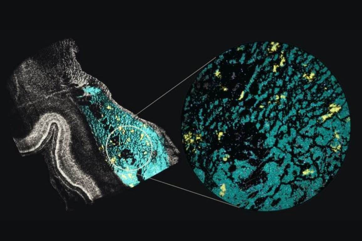
In a landmark study published in Cell, an NIH BRAIN Initiative consortium co-led by UCLA neuroscientists has mapped the molecular and 3D genomic development of the human basal ganglia from mid-pregnancy through infancy. Analyzing nearly 287,000 individual brain cells using advanced single-nucleus methyl-3C sequencing, researchers tracked how developing neurons migrate from temporary prenatal structures known as the ganglionic eminences.
The atlas revealed that spiny projection neurons mature in a highly coordinated manner early in gestation and carry strong enrichment for genetic variants linked to schizophrenia and bipolar disorder, establishing an unprecedented developmental reference to pinpoint when and where psychiatric risk takes root.
During fetal development, the human brain undergoes an extraordinary cellular migration. Newly born neurons arise in transient, specialized germinal zones and travel long distances across developing tissue to assemble into functional circuits.
Because many of these temporary prenatal structures vanish around the time of birth, capturing the molecular transformations that unfold during this migration has historically been one of neurobiology’s greatest technical challenges.
Source: Neuroscience News — Read the full article on the original site ↗